


























































Silent Pressure: Unmasking the Chronic Venous Insufficiency Epidemic and Modern Pathways to Care

Leg swelling after a long day is easy to dismiss as mere fatigue. The same is true of aching calves, ankle discoloration, or varicose veins that have slowly become more prominent over the years. Yet, when these symptoms return day after day, they are rarely benign. Instead, they often point to a progressive circulatory disorder: Chronic Venous Insufficiency (CVI).
CVI develops when the veins in the lower extremities lose their ability to return blood to the heart efficiently. Rather than flowing upward, blood pools in the lower legs, escalating pressure within the venous system and surrounding tissues. Left unaddressed, this silent accumulation of pressure can lead to debilitating skin changes and chronic, non-healing wounds.
1. Main Facts: The Pathophysiology and Presentation of CVI
At the core of chronic venous insufficiency is a failure of venous hemodynamics. Unlike arteries, which rely on the pumping force of the heart to distribute oxygenated blood, the venous system of the lower limbs must fight gravity to return deoxygenated blood to the heart. To achieve this, the body relies on two primary mechanisms: one-way bicuspid valves inside the veins and the contraction of the calf muscles, often referred to as the "peripheral heart."
[Healthy Veins]                          [Incompetent Veins (CVI)]
   To Heart                                      To Heart
      ^                                             ^
      |                                             |
   |  |  |  <-- Valves Close                     |  |  |  <-- Weakened Valves
   |  x  |      To Prevent                       | /  |      Fail to Close
   |  |  |      Backward Flow                    |v   v|
                                                 |  |  |  <-- Blood Flows Backward
                                                 |  v  |      (Reflux) & Pools
In a healthy circulatory system, walking contracts the calf muscles, squeezing the deep veins and forcing blood upward. As the muscles relax, the one-way valves close, preventing gravity from pulling the blood back down.
In patients with CVI, these valves become damaged, stretched, or incompetent. This impairment allows blood to flow backward—a phenomenon known as venous reflux. As blood pools in the superficial and deep venous systems, venous pressure rises, leading to venous hypertension.
Early vs. Advanced Symptomatology
The clinical presentation of CVI spans a wide spectrum. In its infancy, the condition manifests as subtle, easily ignored discomforts:
- A persistent feeling of heaviness or fatigue in the lower legs, particularly by late afternoon.
- Mild swelling (edema) around the ankles, often noticed when shoes or socks feel unusually tight at the end of the day.
- Localized aching or throbbing that improves when walking or elevating the legs.
As venous hypertension persists, it damages the microcirculation of the skin. This leads to advanced complications:
- Stasis Dermatitis: An inflammatory skin condition characterized by itching, redness, scaling, and weeping fluid, which can easily be mistaken for eczema.
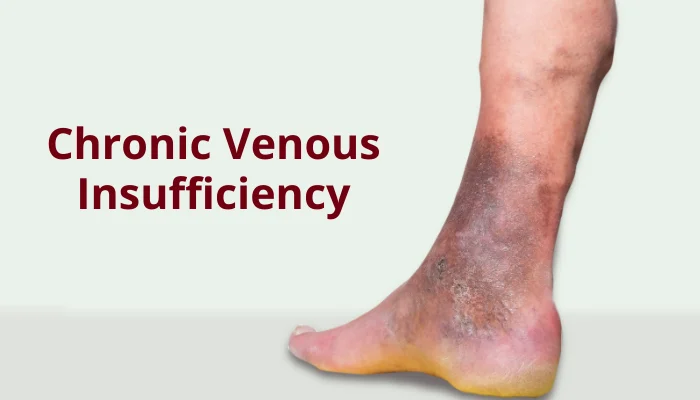
- Hyperpigmentation: A distinctive brownish-rusty discoloration of the skin, typically around the inner ankle. This occurs when high pressure forces red blood cells out of the capillaries into the surrounding tissue, where they break down and leave iron (hemosiderin) deposits in the skin.
- Lipodermatosclerosis: Chronic inflammation that leads to the scarring and hardening of the subcutaneous fat and skin. Over time, this constricts the lower leg, giving it an "inverted champagne bottle" appearance.
- Venous Stasis Ulcers: The most severe consequence of CVI. These shallow, highly painful, and moist wounds usually form near the bony prominence of the inner ankle (medial malleolus). Because the underlying circulation is compromised, these ulcers lack the nutrient-rich blood flow required for natural healing, often remaining open for months or years.
2. Chronology: The Progressive Stages of Venous Disease
Chronic venous insufficiency does not develop overnight. It is a progressive disease that advances through predictable clinical stages. To standardize diagnosis and track this progression, vascular specialists worldwide utilize the CEAP (Clinical, Etiologic, Anatomic, Pathophysiologic) classification system.
The clinical component of this system (C0 through C6) offers a clear chronology of how venous disease advances when left untreated:
The CEAP Chronological Stages
| CEAP Stage | Clinical Classification | Description & Pathological Progression |
|---|---|---|
| C0 | No visible signs | No objective signs of venous disease are visible or palpable, though patients may complain of subjective symptoms like leg heaviness or aching. |
| C1 | Telangiectasias or reticular veins | Commonly known as spider veins (under 1 mm) or blue feeding veins (1–3 mm). These represent early microvascular changes and elevated localized pressure. |
| C2 | Varicose veins | Dilated, tortuous superficial veins measuring 3 mm or larger. At this stage, superficial venous valves have failed, causing visible bulging. |
| C3 | Edema | Persistent swelling of the lower leg and ankle, driven by fluid escaping the high-pressure venous capillaries into the interstitial tissue. |
| C4 | Skin and subcutaneous changes | Divided into C4a (pigmentation or eczema) and C4b (lipodermatosclerosis or atrophie blanche). The skin becomes thin, fragile, discolored, and scarred. |
| C5 | Healed venous ulcer | A history of open wounds that have successfully closed, though the underlying skin remains highly vulnerable to recurrent breakdown. |
| C6 | Active venous ulcer | An open, active wound. This stage represents end-stage venous insufficiency, requiring intensive wound care and vascular intervention. |
3. Supporting Data: Risk Factors and Epidemiological Realities
Epidemiological data reveals that venous disease is highly prevalent, though its exact incidence is often misrepresented. Some public health campaigns claim that up to 40% of the U.S. population suffers from advanced chronic venous insufficiency. However, clinical studies show that while milder forms of venous disease (such as spider veins and uncomplicated varicose veins, CEAP C1–C2) are indeed widespread, advanced CVI (CEAP C3–C6) affects a smaller, yet highly significant, portion of the population—estimated at roughly 2% to 5% of adults.
[Venous Disease Spectrum in the Adult Population]

  [=========================================] Milder Venous Signs (C1-C2)
  ~30% - 40% of Adults (Spider/Varicose Veins)

  [=====] Advanced CVI (C3-C6)
  ~2% - 5% of Adults (Chronic Edema, Skin Changes, Active Ulcers)
The development of CVI is rarely tied to a single cause. Instead, it is driven by a combination of predisposing risk factors:
- Deep Vein Thrombosis (DVT) and Post-Thrombotic Syndrome (PTS): A history of DVT is one of the most critical risk factors. When a blood clot forms in the deep veins, it can permanently destroy the delicate valves. Even after the clot dissolves, the damaged valves allow blood to flow backward, a condition known as post-thrombotic syndrome, which frequently accelerates the onset of severe CVI.
- Gender and Hormonal Influences: Women are diagnosed with venous insufficiency more frequently than men. This disparity is largely driven by pregnancy. During pregnancy, circulating progesterone relaxes the smooth muscle walls of the veins, while increased blood volume and the physical pressure of the growing uterus on the pelvic veins restrict venous return. While symptoms often improve postpartum, successive pregnancies can cause cumulative, permanent damage to venous valves.
- Age and BMI: The risk of CVI increases with age as vein walls naturally lose elasticity and muscle mass declines. Obesity (BMI $ge$ 30) further compounds this risk by dramatically increasing intra-abdominal pressure, which acts as a physical barrier to blood traveling upward from the lower limbs.
- Occupational Inactivity: Individuals who spend long periods standing or sitting without walking—such as assembly line workers, teachers, cashiers, and office desk workers—are at elevated risk. Without regular calf muscle contraction, venous blood pools in the lower extremities, continuously stressing the venous valves.
4. Official Responses: Diagnostic Standards and Clinical Guidelines
Major medical institutions, including the Society for Vascular Surgery (SVS), the American Venous Forum (AVF), and the University of Rochester Medical Center, emphasize that CVI requires objective diagnostic confirmation before initiating advanced therapies.
The Diagnostic Gold Standard: Duplex Ultrasound
A physical examination is highly informative, but the definitive diagnosis of CVI relies on Duplex Ultrasound. This non-invasive test combines traditional anatomical ultrasound imaging with Doppler technology to evaluate both the structure of the veins and the speed and direction of blood flow.
During a diagnostic ultrasound:
- The patient is typically examined in a standing position, which allows gravity to exert downward pressure on the blood column, making venous reflux easier to detect.
- The sonographer measures the time it takes for blood to flow backward when pressure is applied and released (reflux time). A reflux time greater than 0.5 seconds in superficial veins, or greater than 1.0 second in deep veins, is clinically accepted as diagnostic of venous insufficiency.
- The scan maps out precisely which vein segments are incompetent, distinguishing between superficial vein reflux (e.g., the great saphenous vein) and deep vein reflux.
[Normal Flow]                            [Reflux Detected]
  Squeeze Calf -> Blood travels UP          Squeeze Calf -> Blood travels UP
  Release Calf -> Flow stops immediately    Release Calf -> Blood flows BACKWARD
                                            (Reflux time > 0.5s = Incompetence)
Differential Diagnosis and Safety Screens
Before prescribing treatment—particularly compression therapy—clinical guidelines require physicians to rule out other systemic causes of bilateral leg swelling, such as congestive heart failure, renal failure, liver cirrhosis, or lymphedema.
Crucially, clinicians must assess the patient’s arterial circulation using an Ankle-Brachial Index (ABI) test. Applying tight compression garments to a limb with unrecognized Peripheral Artery Disease (PAD) can severely restrict arterial blood flow, risking tissue necrosis or limb loss. As a standard safety protocol, compression therapy is contraindicated or must be heavily modified if the patient’s ABI is below 0.8.
5. Treatment Options: A Multidisciplinary Approach to Care
The management of CVI is highly customized, shifting from conservative lifestyle interventions to advanced minimally invasive procedures depending on the patient’s CEAP stage and ultrasound findings.
Conservative and Lifestyle Therapies
For early-stage CVI, conservative measures remain the cornerstone of therapy:
- Graduated Compression Therapy: Compression stockings apply the highest pressure at the ankle, gradually decreasing up the leg. This external support counteracts venous hypertension, reduces swelling, and helps push pooled blood back into the deep circulation. Stockings must be fitted carefully and are typically worn during waking hours and removed at night.
- Calf Muscle Activation: Regular physical activity, particularly walking, is vital. It engages the calf muscle pump to actively empty the lower limb veins.
- Strategic Leg Elevation: Patients are advised to elevate their legs above the level of their heart for 20 to 30 minutes, three to four times a day. This utilizes gravity to naturally drain fluid and reduce venous pressure.
          [Elevation Principle]

                 (Heart Level)
                     O  <============ [Normal Venous Return]
                    /|
                    /    _.._  <==== [Legs elevated above heart]
                         (____)
Minimally Invasive Interventions
When conservative therapies fail to control symptoms, or when advanced complications like non-healing ulcers develop, modern medicine offers highly effective, outpatient procedures designed to eliminate the source of reflux.
====================================================================
Procedure Type          Mechanism of Action             Recovery Time
====================================================================
Endovenous Thermal      Uses radiofrequency or laser    1 to 2 days
Ablation (RFA/EVLO)     heat to collapse the damaged
                        vein from the inside.

Sclerotherapy           Injects a chemical sclerosant   Immediate
                        to seal spider or small varicose
                        veins.

VenaSeal (Adhesive)     Seals the diseased vein using   Immediate (No
                        medical-grade glue, avoiding    thermal energy)
                        heat or nerve injury risks.

Vein Stripping          Surgically removes the vein.     2 to 4 weeks
(Traditional)           Reserved for highly complex
                        anatomies.
====================================================================- Endovenous Thermal Ablation: Utilizing either Radiofrequency Ablation (RFA) or Endovenous Laser Treatment (EVLT), a physician inserts a thin catheter into the diseased vein under ultrasound guidance. Local anesthetic is applied around the vein, and thermal energy is delivered to heat and damage the vein wall, causing it to collapse and seal shut. The body naturally diverts blood flow to neighboring healthy veins.
- Non-Thermal Ablation (VenaSeal): This modern technique utilizes a medical-grade cyanoacrylate adhesive (glue) to seal the diseased vein. Because it does not use heat, it eliminates the risk of nerve injury and does not require extensive local anesthesia.
- Sclerotherapy: Primarily used for smaller varicose veins and spider veins, this procedure involves injecting a liquid or foam sclerosant directly into the vein, causing it to spasm, scar, and dissolve over several weeks.
- Traditional Vein Surgery: While minimally invasive techniques have largely replaced open surgery, procedures such as vein ligation and stripping are still utilized for highly complex vascular structures or when previous catheter-based interventions have failed.
6. Implications: The Economic, Societal, and Future Outlook of CVI
The societal and economic impact of chronic venous insufficiency is profound. Venous ulcers, which affect roughly 1% of the adult population at some point in their lives, carry an immense financial burden.
In the United States alone, the annual cost of treating chronic venous ulcers is estimated to exceed $2.5 billion. This cost is driven by frequent physician visits, specialized dressings, home health care, and the management of secondary infections like cellulitis.
[The Vicious Cycle of Advanced CVI]

   Venous Hypertension
        │
        ▼
   Skin Breakdown (Ulceration) ──► High Healthcare Costs ($2.5B+ annually)
        │
        ▼
   Reduced Mobility & Work Absences ──► Loss of Productivity
Beyond the financial toll, CVI significantly diminishes quality of life. The chronic pain, constant fluid drainage, and restricted mobility associated with advanced venous ulcers often lead to social isolation, depression, and substantial lost productivity in the workforce.
The Impact of the Modern Workplace
The modern shift toward sedentary, office-based work environments has intensified the prevalence of venous disease. Public health experts increasingly emphasize the importance of vascular hygiene in the workplace.
Simple, low-cost interventions—such as incorporating sit-stand desks, encouraging short walking breaks every hour, and promoting simple ankle pump exercises during long flights or desk sessions—are vital steps in preventing the onset of venous reflux.
The Future of Venous Care
The future of CVI management lies in early detection and public education. For decades, swelling and varicose veins were dismissed as cosmetic concerns or inevitable signs of aging. Today, the medical community recognizes that early diagnosis through duplex ultrasound, combined with timely, minimally invasive intervention, can arrest the progression of CVI before irreversible tissue damage, such as lipodermatosclerosis or ulceration, occurs.
Through increased public awareness and proactive medical management, the severe complications of this progressive circulatory disorder can be successfully prevented, allowing patients to maintain their mobility and quality of life for years to come.