


























































The Silent Progression of Chronic Venous Insufficiency: Clinical Realities, Diagnosis, and Modern Care

For millions of people, a long day on their feet ends with a familiar discomfort: heavy, aching legs and ankles that look slightly swollen. It is easy to dismiss these symptoms as the simple price of a hard day’s work. However, when these symptoms persist, recur daily, or are accompanied by changes in skin color and prominent varicose veins, they point to a underlying circulatory issue known as Chronic Venous Insufficiency (CVI).
CVI is a progressive circulatory condition that occurs when the veins in the lower extremities are unable to return blood to the heart efficiently. Rather than a benign cosmetic concern, CVI represents a significant vascular disease that can lead to debilitating complications, including chronic open wounds, if left unmanaged.
1. Main Facts: Understanding the Mechanics of CVI
To understand Chronic Venous Insufficiency, one must look at the mechanics of the circulatory system. While the heart pumps oxygen-rich blood down to the lower extremities through high-pressure arteries, returning that blood back up to the chest against the force of gravity is a complex physiological challenge.
[Healthy Veins]                           [Venous Insufficiency]
  ▲  Upward Flow                             ▲  Upward Flow
  │                                          │
 ───  Valves Close                          ─ ─  Valves Fail to Close
  ▲   Preventing Backflow                    ▼   Reflux (Blood Pools)
  │                                          │
The body relies on two primary mechanisms to accomplish this:
- One-Way Venous Valves: Tiny, delicate bivalve flaps located at regular intervals inside the veins. These valves open to allow blood to flow upward and close immediately to prevent gravity from pulling the blood backward.
- The Calf Muscle Pump: Often referred to by vascular specialists as the "peripheral heart." When a person walks, the contraction of the calf muscles squeezes the deep veins of the leg, forcing blood upward through the open valves.
In patients with CVI, this system breaks down. The venous walls lose their elasticity, causing the veins to stretch and widen. As a result, the valve leaflets can no longer meet in the center to close properly. This structural failure leads to venous reflux—a state where blood flows backward and pools in the lower legs. This pooling raises the hydrostatic pressure within the veins, a pathological state called venous hypertension, which eventually damages the surrounding tissues, microcirculation, and skin.
2. Chronology: From Minor Fatigue to Chronic Ulceration
The clinical progression of CVI is notoriously gradual, often spanning years or even decades. Patients frequently adapt to their symptoms, normalizing their increasing discomfort until the disease reaches an advanced stage.
Early Stage                 Intermediate Stage           Advanced Stage
[Tiredness & Edema]  ───►  [Skin Discoloration]  ───►  [Lipodermatosclerosis]  ───►  [Venous Ulcers]
(Reversible swelling)       (Hemosiderin staining)      (Irreversible scarring)       (Open, chronic wounds)
The Early Phase: Subclinical Symptoms
In the initial stages, the symptoms are subjective and easily ignored. Patients report a vague feeling of heaviness, tiredness, or aching in their calves, particularly by late afternoon or after prolonged sitting or standing.
- Ankle Edema: Mild swelling around the ankles may develop by the end of the day, causing shoes to feel uncomfortably tight.
- Night Cramps and Restlessness: Some patients experience nocturnal calf cramps or restless sensations in their legs, which are often misattributed to electrolyte imbalances or aging.
The Intermediate Phase: Structural and Visual Changes
As venous hypertension persists, fluid and red blood cells leak out of the high-pressure capillaries into the surrounding subcutaneous tissue.
- Corona Phlebectatica: Small, fan-shaped clusters of intradermal veins become visible around the ankle and foot, serving as an early clinical warning of sustained venous hypertension.
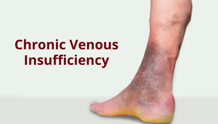
- Hyperpigmentation (Hemosiderin Staining): As red blood cells break down in the skin, the iron-rich pigment hemosiderin deposits into the tissue, turning the skin around the lower calves and ankles a rusty brown or dark grey color.
- Stasis Dermatitis: The skin becomes inflamed, red, dry, itchy, and scaly, frequently mimicking eczema.
The Advanced Phase: Tissue Destruction
Without intervention, chronic inflammation permanently alters the architecture of the lower leg.
- Lipodermatosclerosis: The subcutaneous fat becomes chronically inflamed and progressively replaced by scar tissue. The skin hardens, tightens, and contracts, giving the lower leg a distinctive "inverted champagne bottle" appearance, where the calf remains normal but the ankle becomes narrow and woody.
- Atrophie Blanche: Smooth, ivory-white scars surrounded by dilated capillaries appear on the ankle, representing areas of localized tissue death.
- Venous Stasis Ulcers: The ultimate stage of untreated CVI. Even minor trauma—such as a scratch or a bug bite—can trigger a breakdown of the fragile, poorly oxygenated skin. This results in shallow, highly painful, moist wounds, typically located near the medial malleolus (the inner ankle bone), which can remain open for months or years.
3. Supporting Data and Clinical Metrics
Chronic venous disease is incredibly widespread, though its exact prevalence is frequently misstated. While minor venous issues like spider veins (telangiectasias) and mild varicose veins affect a vast portion of the global adult population, advanced CVI is a distinct, more severe clinical entity.
Risk Factor Analysis
The development of CVI is multifactorial. Key risk factors include:
- History of Deep Vein Thrombosis (DVT): This is one of the most significant clinical precursors. A blood clot in the deep veins can permanently scar and destroy venous valves, leading to a severe form of CVI known as Post-Thrombotic Syndrome (PTS).
- Biological Sex: Women are diagnosed with venous disease significantly more often than men. This is driven by hormonal factors (progesterone relaxes vein walls) and pregnancy, which increases circulating blood volume while the growing uterus mechanically compresses the inferior vena cava, elevating leg vein pressure.
- Occupational Stasis: Professions requiring prolonged, stationary standing or sitting (such as nurses, factory workers, teachers, and office workers) drastically reduce calf muscle pump activation, accelerating venous pooling.
- Obesity and Height: Excess body weight increases intra-abdominal pressure, resisting the upward flow of venous blood. Taller individuals are also at higher risk due to the greater hydrostatic pressure exerted by a taller column of blood.
The CEAP Classification System
To standardize the diagnosis and tracking of chronic venous disorders, vascular specialists worldwide use the CEAP system (Clinical, Etiologic, Anatomic, Pathophysiologic). The clinical portion of this scale provides a clear framework for staging the disease:
| CEAP Stage | Clinical Presentation | Pathological Implication |
|---|---|---|
| C0 | No visible or palpable signs of venous disease | Subjective symptoms (heaviness, aching) may still be present. |
| C1 | Telangiectasias (spider veins) or reticular veins | Superficial, localized venous dilation; primarily cosmetic. |
| C2 | Varicose veins (distended, tortuous veins ≥ 3mm) | Indicates valve failure in the superficial venous system. |
| C3 | Edema (swelling of the lower leg and ankle) | Fluid accumulation due to elevated capillary pressure. |
| C4a | Pigmentation or eczema (stasis dermatitis) | Inflammatory changes and red blood cell extravasation. |
| C4b | Lipodermatosclerosis or atrophie blanche | Deep tissue scarring, hardening, and localized necrosis. |
| C5 | Healed venous ulcer | Fragile, scarred skin at high risk of re-ulceration. |
| C6 | Active venous ulcer | Open, infected, or exudative wound requiring intensive wound care. |
4. Official Responses and Expert Clinical Guidelines
Leading vascular organizations, including the Society for Vascular Surgery (SVS), the American Venous Forum (AVF), and academic centers like the University of Rochester Medical Center and UCSF Vascular Surgery, have established clear diagnostic and treatment guidelines to optimize patient outcomes.
       [Symptomatic Patient]
                 │
        [Duplex Ultrasound]
                 │
       ┌─────────┴─────────┐
       ▼                   ▼
[Reflux Confirmed]   [Reflux Absent]
       │                   │
[Conservative Care]  [Investigate Other Causes]
(Compression, Elev.) (Heart, Kidney, Lymphedema)
       │
[Refractory / Advanced CVI?]
       │
       ▼
[Minimally Invasive Procedures]
(Ablation, Sclerotherapy)
The Gold Standard of Diagnosis: Duplex Ultrasound
Vascular experts agree that a physical exam alone is insufficient to diagnose CVI or plan treatment.
- Duplex Ultrasound is the diagnostic gold standard. It combines traditional anatomical imaging with color Doppler technology to measure the direction and velocity of blood flow.
- A diagnosis of venous reflux is confirmed when the ultrasound demonstrates retrograde (backward) blood flow lasting longer than 0.5 seconds in the superficial veins, or longer than 1.0 second in the deep venous system.
- For complex cases—such as suspected pelvic congestion syndrome or anatomical obstructions in the abdomen (e.g., May-Thurner Syndrome)—advanced cross-sectional imaging like CT Venography or Magnetic Resonance Venography (MRV) is utilized.
Diagnostic Rule-Outs and Safety Protocols
Experts emphasize that leg swelling is a non-specific symptom. Clinicians must rule out other systemic causes of bilateral leg edema, such as:
- Congestive heart failure (CHF)
- Chronic kidney disease (CKD)
- Hepatic cirrhosis
- Lymphedema (which can coexist with venous disease, termed phlebolymphedema)
Crucial Safety Rule: Before initiating any form of compression therapy, a patient’s arterial circulation must be evaluated. Applying tight compression stockings to a patient with unrecognized Peripheral Artery Disease (PAD) can severely restrict arterial blood flow, leading to tissue ischemia or gangrene. Specialists often perform an Ankle-Brachial Index (ABI) test; if the ABI is below 0.8, high-pressure compression is contraindicated.
5. Implications: The Societal Burden and the Evolution of Modern Treatment
The implications of CVI extend far beyond individual discomfort. Chronic venous ulcers represent a massive economic and clinical burden on global healthcare systems, costing billions of dollars annually in wound dressings, nursing care, and lost productivity. Fortunately, the treatment paradigm has shifted dramatically over the past two decades.
The Shift in Treatment Paradigms
Historically, the primary surgical option for severe varicose veins and CVI was "vein stripping"—an invasive surgical procedure performed under general anesthesia that involved physically pulling the saphenous vein out of the leg. This required a lengthy recovery time and carried a high risk of nerve damage.
Today, traditional stripping has been largely replaced by outpatient, minimally invasive techniques:
───────────────────────────────────────────────────────────────────────────
Therapy Category         Method                       How It Works
───────────────────────────────────────────────────────────────────────────
Thermal Ablation         Radiofrequency / Laser       Heat is applied via a catheter
                                                      to collapse and seal the vein.

Non-Thermal Ablation     VenaSeal (Adhesive)          Medical-grade glue is used to
                                                      instantly close the diseased vein.

Chemical Treatment       Sclerotherapy                An sclerosant agent is injected,
                                                      irritating and closing the vein.
───────────────────────────────────────────────────────────────────────────
Once the diseased superficial vein is closed, the body naturally reroutes blood flow through healthier, deeper venous pathways, instantly lowering the venous pressure in the lower leg.
Conservative Management and Prevention
For patients in the early stages (C0 to C3), or those who are not candidates for procedures, conservative lifestyle management remains the cornerstone of care:
- Graduated Compression Stockings: These garments provide a tight squeeze at the ankle that gradually decreases up the leg, physically forcing pooled fluid out of the tissues and back into the venous circulation.
- Structured Exercise: Regular walking, swimming, or cycling strengthens the calf muscles, enhancing the efficiency of the calf muscle pump.
- True Leg Elevation: To successfully reduce venous pressure, patients must elevate their legs above the level of the heart for 20 to 30 minutes, 3 to 4 times a day. Merely resting feet on a footstool is insufficient to counter the forces of gravity.
Red Flag Symptoms Requiring Urgent Intervention
While CVI is generally a slow, chronic disease, certain acute developments require immediate emergency evaluation:
- Sudden, Unilateral Swelling: If one leg swells rapidly over a few hours, it may indicate an acute Deep Vein Thrombosis (DVT).
- Chest Pain or Shortness of Breath: These are classic signs of a Pulmonary Embolism (PE), a life-threatening emergency occurring when a DVT breaks loose and travels to the lungs.
- Signs of Infection (Cellulitis): Rapidly spreading redness, warmth, severe pain, or pus draining from a venous ulcer or skin break.
- Acute Hemorrhage: Thin, high-pressure varicose veins near the skin surface can rupture and bleed profusely. This requires immediate leg elevation and direct, firm pressure.
Patient Advocacy: Questions to Ask Your Specialist
Patients experiencing chronic leg symptoms should actively participate in their care by asking key diagnostic and therapeutic questions during their medical appointments:
- "Did my duplex ultrasound show reflux in the deep veins, the superficial veins, or both?"
- "Is my arterial circulation strong enough to safely wear prescription compression stockings?"
- "Are my skin changes reversible, or has permanent scarring (lipodermatosclerosis) occurred?"
- "Am I a candidate for a minimally invasive office procedure, or should I start with conservative therapy?"
- "If I have a history of blood clots, how does that change my long-term treatment plan?"
Conclusion
Chronic Venous Insufficiency is a progressive, structural vascular disease that requires proactive management. By understanding that heavy, swollen legs are a sign of underlying circulatory dysfunction rather than a normal consequence of aging or fatigue, patients can seek early evaluation. Through a combination of diagnostic ultrasound, conservative compression therapy, and modern, minimally invasive procedures, the progressive damage of CVI can be halted, protecting both the skin and the patient’s long-term mobility.