


























































The Silent Progression of Chronic Venous Insufficiency: Clinical Realities, Diagnostic Paths, and Modern Therapeutics

Leg swelling after a long day is easy to dismiss as simple fatigue. The same is true of aching calves, ankle discoloration, or varicose veins that have slowly become more prominent over the years. However, when these issues return day after day, they often point to a underlying medical condition: Chronic Venous Insufficiency (CVI).
CVI is a progressive circulatory disorder that arises when the veins in the lower extremities are unable to return blood to the heart efficiently. This leads to blood pooling in the lower legs, escalating pressure within the venous system, and causing microvascular changes that damage the surrounding skin and subcutaneous tissues. While early symptoms may seem minor, untreated CVI can progress to severe, life-altering complications, including chronic pain, permanent skin changes, and debilitating open wounds known as venous ulcers.
Main Facts: The Pathophysiology of Venous Insufficiency
To understand chronic venous insufficiency, one must first understand the anatomy of venous return. Unlike arteries, which rely on the pumping force of the heart to distribute oxygenated blood throughout the body, the venous system of the lower extremities must fight gravity to return deoxygenated blood back to the heart.
[Heart] <------------------------------- (Deoxygenated Blood)
   ^
   |  (Deep Veins / Superficial Veins)
   |
[Healthy One-Way Valves]  <--- Prevent Backward Flow (Reflux)
   ^
   |  (Calf Muscle Pump Activates During Walking)
   |
[Lower Extremities]
To accomplish this, the body utilizes two primary mechanisms:
- One-Way Venous Valves: Tiny, bicuspid valves situated along the interior walls of veins. These valves open to allow blood to flow upward toward the heart and snap shut to prevent gravity from pulling blood backward (reflux).
- The Calf Muscle Pump: Often referred to by vascular specialists as the "peripheral heart." When a person walks, the contraction of the calf muscles compresses the deep veins of the leg, squeezing blood upward.
In a patient with CVI, this system breaks down. The venous walls can lose their elasticity, causing the veins to stretch and widen. As a result, the delicate valve leaflets no longer meet in the center, allowing blood to flow backward and pool in the lower leg. This backflow is medically termed venous reflux.
As blood pools, the hydrostatic pressure within the veins increases—a state known as venous hypertension. This elevated pressure is transmitted down to the smallest capillaries, forcing fluid, red blood cells, and inflammatory proteins out of the blood vessels and into the surrounding interstitial tissue. This microvascular leakage is the fundamental driver of the swelling, pain, and tissue damage characteristic of advanced CVI.
Distinguishing Varicose Veins from CVI
While varicose veins and CVI are closely linked, they are not synonymous:
- Varicose Veins: These are dilated, twisting, superficial veins visible just beneath the skin. A person can have prominent varicose veins without suffering from deep-system venous insufficiency or chronic swelling.
- Chronic Venous Insufficiency (CVI): This represents a deeper, more systemic failure of the venous system. A patient may present with severe deep-vein valve damage and profound lower-leg swelling without exhibiting any visible varicose veins on the surface.
Chronology: The Progressive Stages of Venous Disease
Chronic venous insufficiency does not develop overnight. It is a slow, degenerative disease that progresses through predictable clinical stages over months, years, or even decades. Understanding this timeline allows clinicians to intervene early before irreversible tissue damage occurs.
[Stage 1: Asymptomatic / Reflux]
       │
       ▼
[Stage 2: Mild Symptomatic] (Heaviness, tight shoes, evening edema)
       │
       ▼
[Stage 3: Visible Vascular Changes] (Spider veins, varicose veins, corona phlebectatica)
       │
       ▼
[Stage 4: Inflammatory Tissue Changes] (Stasis dermatitis, lipodermatosclerosis)
       │
       ▼
[Stage 5: Ulceration] (Active or healed venous stasis ulcers)
Phase 1: Microvascular Insufficiency and Early Reflux (The Asymptomatic Stage)
At the onset, valve degradation occurs silently. The patient may feel entirely healthy, though an ultrasound would reveal early signs of retrograde blood flow in either the superficial or deep venous systems.
Phase 2: Mild Symptomatic Presentation (The Warning Signs)
As venous pressure begins to accumulate, the patient experiences transient symptoms. Typically, these are most pronounced in the late afternoon or evening, especially after prolonged standing or sitting.
- Early Indicators: A sensation of heaviness or fatigue in the calves, mild swelling around the ankles, and shoes that feel uncomfortably tight by the end of the day.
- Relief Patterns: Symptoms temporarily improve when the patient walks (activating the calf muscle pump) or elevates their legs.
Phase 3: Visible Vascular Alterations
Over time, the chronic pressure forces superficial veins to dilate.
- Spider Veins (Telangiectasias): Tiny, damaged capillaries near the skin’s surface.
- Varicose Veins: Larger, bulging, blue or purple veins.
- Corona Phlebectatica: A fan-shaped pattern of small, intradermal veins around the ankle and foot. This is a critical clinical marker indicating that high venous pressure has been sustained for a long period.
Phase 4: Chronic Inflammatory and Skin Changes (Stasis Dermatitis)
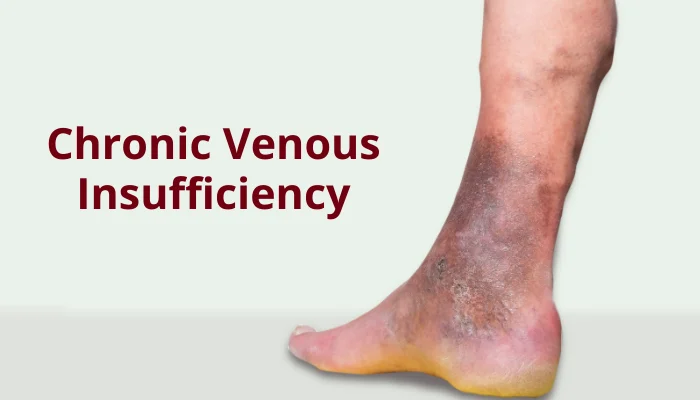
If venous hypertension remains untreated, the leakage of red blood cells into the surrounding tissue leads to the breakdown of hemoglobin. This releases iron into the skin, causing a dark brown or rust-colored discoloration known as hemosiderin staining.
- Stasis Dermatitis: The skin becomes highly inflamed, dry, itchy, and scaly, frequently misdiagnosed as eczema.
- Lipodermatosclerosis: Chronic inflammation leads to scarring of the skin and subcutaneous fat. The tissue hardens, and the lower leg narrows significantly just above the ankle, giving the leg an appearance often described as an "inverted champagne bottle."
Phase 5: Ulceration (End-Stage CVI)
The severely compromised, hardened skin becomes incredibly fragile. Even a minor scratch, bump, or insect bite can cause the skin to break open. Because the underlying circulation is so poor, the body cannot deliver the oxygen and nutrients required for healing. This results in a venous stasis ulcer—a shallow, painful, moist wound, typically located near the bony prominence of the inner ankle (medial malleolus), which can remain open for months or years.
Supporting Data: Risk Factors and Clinical Staging
To standardize the diagnosis and treatment of venous disease, vascular specialists worldwide utilize the CEAP Classification System. This system categorizes the disease based on Clinical signs, Etiology (cause), Anatomy, and Pathophysiology.
The CEAP Classification Scale
| CEAP Stage | Clinical Findings & Characteristics |
|---|---|
| C0 | No visible or palpable signs of venous disease. |
| C1 | Telangiectasias (spider veins) or reticular veins. |
| C2 | Varicose veins (dilated veins $>3$ mm in diameter). |
| C3 | Edema (swelling of the leg and ankle without skin changes). |
| C4 | Changes in the skin and subcutaneous tissue (hyperpigmentation, stasis eczema, lipodermatosclerosis). |
| C5 | Healed venous ulcer. |
| C6 | Active venous ulcer (open, draining wound). |
Epidemiological Data and Risk Factors
Epidemiological studies indicate that venous disorders are highly prevalent, though estimates vary based on the specific CEAP stage studied. While mild venous changes (C1-C2) affect a broad segment of the global adult population, advanced chronic venous insufficiency (C3-C6) is estimated to affect roughly 2% to 5% of the population, with prevalence rising sharply with age.
The development of CVI is rarely tied to a single cause; rather, it is driven by a combination of predisposing risk factors:
                  ┌────────────────────────┐
                  │   Advanced Age (>50)   │
                  └───────────┬────────────┘
                              │
  ┌──────────────────────┐    ▼    ┌──────────────────────┐
  │ Pregnancy/Hormones   ├─────────┤ Previous DVT/Clots   │
  └──────────────────────┘    ▲    └──────────────────────┘
                              │
  ┌──────────────────────┐    ▼    ┌──────────────────────┐
  │ Obesity (Abdominal)  ├─────────┤ Prolonged Immobility │
└──────────────────────┘ └──────────────────────┘- Previous Deep Vein Thrombosis (DVT): A blood clot in the deep veins can permanently scar and destroy venous valves. This specific etiology is known as Post-Thrombotic Syndrome (PTS), which is one of the most severe and difficult-to-treat forms of CVI.
- Pregnancy and Hormonal Fluctuations: Pregnancy introduces a triple threat to the venous system: circulating progesterone relaxes the smooth muscle in vein walls, blood volume increases significantly, and the expanding uterus physically compresses the vena cava, restricting venous return from the legs.
- Obesity: Excess abdominal fat increases intra-abdominal pressure, which acts as a physical barrier to blood traveling upward from the femoral veins.
- Occupational Immobility: Professions that require hours of uninterrupted standing (e.g., nurses, teachers, factory workers) or sitting (e.g., office workers, long-haul drivers) deprive the body of the calf muscle pump, leading to prolonged venous stasis.
- Height: Taller individuals have a higher baseline hydrostatic pressure in their lower extremities simply due to the physical distance blood must travel against gravity.
Official Responses: Clinical Guidelines and Diagnostic Standards
Major medical institutions, including the Society for Vascular Surgery (SVS), the American Venous Forum (AVF), and the University of Rochester Medical Center, emphasize that a formal diagnosis is essential before any therapeutic intervention begins. Leg swelling is a non-specific symptom that can point to systemic diseases, and incorrect treatment can be dangerous.
The Diagnostic Gold Standard: Duplex Ultrasound
The cornerstone of CVI diagnosis is the Duplex Ultrasound. This non-invasive, painless test combines traditional B-mode ultrasound imaging (to visualize the structure of the veins and look for old blood clots) with Doppler technology (to measure the direction and velocity of blood flow).
During a diagnostic ultrasound:
- The patient is typically examined in a standing position, which allows gravity to exert natural downward pressure on the blood columns.
- The sonographer performs manual compression maneuvers (squeezing the calf) to force blood upward, then observes whether the blood flows backward when the squeeze is released.
- Reflux is quantified by time: In the deep veins, backward flow lasting longer than 1.0 second is considered abnormal; in the superficial veins, the threshold for abnormal reflux is greater than 0.5 seconds.
[Calf Squeeze] ---------> Blood moves upward (Normal)
[Squeeze Released] -----> Normal Valve: Blood stops immediately (<0.5s)
                   -----> Damaged Valve (Reflux): Blood flows backward (>0.5s)
Differential Diagnosis and Safety Precautions
Medical guidelines stress the importance of ruling out other systemic causes of bilateral leg swelling, such as:
- Congestive heart failure (CHF)
- Chronic kidney disease or renal failure
- Hepatic cirrhosis
- Lymphedema
- Medication-induced edema (particularly calcium channel blockers used for hypertension)
The Critical Link to Peripheral Artery Disease (PAD)
Before initiating any form of compression therapy, clinicians must assess the patient’s arterial circulation. Applying tight compression stockings to a leg that has compromised arterial blood flow can completely cut off perfusion, leading to acute ischemia, tissue necrosis, or amputation.
To prevent this, guidelines recommend performing an Ankle-Brachial Index (ABI) test. If the ABI is less than 0.5 to 0.8, standard high-pressure compression therapy is strictly contraindicated.
Implications: Treatment Paradigms, Prevention, and Urgent Indicators
The management of chronic venous insufficiency has evolved from highly invasive surgeries toward conservative therapies and outpatient, minimally invasive procedures.
                     ┌───────────────────────────┐
                     │    Conservative Care      │
                     │  (Compression, Elevation, │
                     │   Exercise, Skin Care)    │
                     └─────────────┬─────────────┘
                                   │
                    If symptoms persist / Ulcer forms
                                   │
                                   ▼
                     ┌───────────────────────────┐
                     │ Minimally Invasive Tech   │
                     │  (Endovenous Ablation,    │
                     │      Sclerotherapy)       │
                     └─────────────┬─────────────┘
                                   │
                       If anatomy is complex
                                   │
                                   ▼
                     ┌───────────────────────────┐
                     │    Traditional Surgery    │
                     │  (Vein Ligation/Stripping)│
                     └───────────────────────────┘
Conservative Treatment Modalities
Conservative therapy remains the first line of defense for almost all stages of CVI.
- Graduated Compression Therapy: Elastic stockings designed to apply the tightest pressure at the ankle, gradually decreasing as the stocking moves up the calf. This external pressure counteracts venous hypertension, reduces edema, and helps push fluid back into the lymphatic and venous vessels.
- Structured Exercise (Walking): Regular walking programs strengthen the calf muscles, directly improving the efficiency of the calf muscle pump.
- Strategic Leg Elevation: Patients are advised to elevate their legs above the level of their heart for 15 to 30 minutes, 3 to 4 times a day. Merely resting one’s feet on a footstool is insufficient; the ankles must be physically higher than the atrium of the heart to allow gravity to drain the pooled fluid.
- Advanced Skin Care: For patients with stasis dermatitis, keeping the skin clean, dry, and highly moisturized with barrier creams is vital to prevent microscopic skin tears that can lead to bacterial infections (cellulitis).
Minimally Invasive Interventions
If conservative measures fail to manage symptoms, or if a patient presents with an active venous ulcer (CEAP C6), modern endovascular procedures can target and eliminate the refluxing veins.
- Endovenous Thermal Ablation (Radiofrequency or Laser): Under local anesthesia, a thin catheter is inserted into the diseased superficial vein (typically the Great Saphenous Vein). Using either radiofrequency energy or laser heat, the physician seals the vein shut from the inside. The body naturally redirects the blood flow through healthier deep veins, and the closed vein eventually dissolves into scar tissue.
- Sclerotherapy: A chemical sclerosant (in liquid or foam form) is injected directly into smaller varicose veins or reticular veins. The chemical irritates the lining of the blood vessel, causing it to collapse, stick together, and close.
- Non-Thermal Adhesives (Vein Glue): A medical-grade cyanoacrylate adhesive is injected into the diseased vein via a catheter, sealing the vein closed instantly without the need for heat or extensive local anesthesia.
When to Seek Urgent Medical Care
While CVI is generally a chronic, slow-moving condition, there are several acute complications that require emergency medical evaluation:
                                 [Urgently Seek Medical Attention]
                                                │
         ┌──────────────────────────────────────┼──────────────────────────────────────┐
         │                                      │                                      │
         ▼                                      ▼                                      ▼
[Sudden Unilateral Swelling]             [Uncontrolled Bleeding]               [Spreading Redness/Heat]
- Possible Deep Vein Thrombosis          - Ruptured high-pressure varicosity    - Sign of acute Cellulitis
- Risk of Pulmonary Embolism - Requires immediate antibiotics- Sudden, Unilateral Leg Swelling: If one leg swells rapidly while the other remains normal, this is a hallmark sign of an acute Deep Vein Thrombosis (DVT). If a piece of this clot breaks free, it can travel to the lungs, causing a life-threatening Pulmonary Embolism (PE). Immediate emergency care is required if this swelling is accompanied by sudden shortness of breath, chest pain, or coughing up blood.
- Severe, Uncontrolled Bleeding: Because varicose veins are under high hydrostatic pressure, a minor nick or rupture of an overlying thin layer of skin can result in profuse bleeding. If this occurs, the patient must lie down immediately, elevate the leg straight up above the heart, apply firm, direct pressure, and seek emergency care if the bleeding does not stop.
- Spreading Redness, Heat, and Fever: These are signs of cellulitis, a deep bacterial skin infection that can rapidly spread through tissues and enter the bloodstream (sepsis) if not treated promptly with antibiotics.
Questions Patients Should Ask Their Doctor
For patients experiencing persistent leg swelling, heaviness, or skin changes, preparing the right questions for their vascular consultation can ensure they receive a comprehensive care plan:
- Does my duplex ultrasound show reflux in my deep veins, superficial veins, or both?
- Is my ankle-brachial index (ABI) normal, and is it safe for me to wear compression stockings?
- Are my symptoms related to a past blood clot (Post-Thrombotic Syndrome)?
- What level of graduated compression (measured in mmHg) is appropriate for my stage of disease?
- Am I a candidate for a minimally invasive, outpatient procedure like endovenous ablation to resolve my venous reflux?
- What lifestyle modifications can I implement at work and home to slow the progression of this disease?
Living with CVI: The Long-Term Outlook
Chronic venous insufficiency is a lifelong vascular condition, but it is highly manageable. With early detection, consistent use of compression therapy, regular movement, and timely medical interventions, patients can prevent the debilitating progression from mild swelling to chronic ulceration, maintaining their mobility and overall quality of life.